Cerebrospinal Fluid Remains Best Diagnostic Method
Written by |
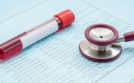


Examining cerebrospinal fluid — the liquid surrounding the brain and spinal cord — remains the best way to diagnose aromatic L-amino acid decarboxylase (AADC) deficiency, a recent study confirms.
The investigation also found that measuring treatment response by routinely re-examining this fluid appears unnecessary, and that certain molecules found in the blood might prove useful as biomarkers, although more studies are needed to confirm this.
The study, “Blood, urine and cerebrospinal fluid analysis in TH and AADC deficiency and the effect of treatment,” was published in the journal Molecular Genetics and Metabolism Reports.
The AADC enzyme is needed to produce serotonin and dopamine, two neurotransmitters, which are molecules used to transmit nervous signals from the brain to the rest of the body.
Measuring the amounts of intermediate molecules in the production of these neurotransmitters — their metabolites — in the cerebrospinal fluid (CSF) is a tried-and-true way to diagnose AADC deficiency.
Since collecting CSF requires an invasive spinal tap, blood and urine are two attractive alternatives to diagnose AADC. However, the diagnostic value of neurotransmitter metabolite levels in these bodily fluids remains poorly studied.
Is also is unclear how neurotransmitter metabolite levels in the CSF change over the course of treatment — and whether they can be used to monitor treatment efficacy.
To better answer these questions, scientists affiliated with Radboud University, in the Netherlands, reviewed patient data and investigated patterns of neurotransmitter metabolism in urine, blood, and CSF before and during treatment.
They also examined prolactin levels in serum (the liquid portion of blood) before and during treatment. Dopamine normally limits the release of this hormone into the bloodstream, leading to the hypothesis that it should be elevated in AADC deficiency. Whether prolactin levels can aid in AADC deficiency diagnosis, however, is unclear.
The researchers collected patient data from published medical literature and from their own hospital’s database.
Among untreated AADC patients, 34 had blood measurements for 3-O-methyldopa (3-OMD, greater levels of which are a potential “red flag” for AADC deficiency). In all cases, 3-OMD levels were strongly elevated.
Conversely, serum serotonin levels were decreased in all seven untreated patients with those measurements. Serum prolactin was elevated in 34 of 37 patients; the remaining three had received treatment and had prolactin at normal levels.
While other blood metabolites could reflect the metabolic problems in only a limited number of AADC patients, the researchers pointed to 3-OMD as a clear exception, demonstrating that measuring this biomarker in dried blood spots as part of newborn screening programs is “interesting and promising to decrease the long diagnostic delay that is often present in this disorder,” they wrote.
Urine dopamine levels were either normal or increased in 21 of 24 measured patients, while the biomarkers 5-hydroxyindoleacetic acid (5-HIAA) was lower in nine of 10 patients, and vanillactic acid was elevated in 19 of 20 patients.
The findings demonstrated that urine levels of neurotransmitter metabolites “should be interpreted with extreme caution and are of very limited diagnostic value.”
Treatment did not appear to affect the levels of these metabolites significantly, except for a rise in homovanillic acid (another biomarker) in the urine and CSF, in response to levodopa therapy.
Levodopa, however, is effective only in a subset of AADC deficiency patients whose mutations make them responsive to it.
The investigators found 16 patients who had received gene therapy for AADC deficiency, 12 of whom had homovanillic acid and 5-HIAA CSF measurements both before and after treatment.
Homovanillic acid increased in eight of these patients, but still remained low in five of them, while 5-HIAA levels remained greatly decreased in nine, and increased in three to moderately or mildly decreased levels.
“In our small and retrospective series, there is no clear correlation between alterations in CSF [neurotransmitter] metabolites and clinical response to medical treatment,” the scientists wrote.
They noted that in the case of gene therapy, homovanillic acid showed a “modest increase” in most patients, yet could remain strongly decreased even when patients showed clear clinical responses.
For this reason, they found it “wiser and more patient friendly” to evaluate gene therapy recipients using clinical parameters, rather than more invasive biochemical measurements.
Although the investigators determined that the value of serum prolactin should be explored further, they concluded that overall, their study “confirms that cerebrospinal fluid is the most informative body fluid to measure monoamine neurotransmitter metabolites when AADC … is suspected, and that routine follow-up of cerebrospinal fluid measurements to estimate treatment response is not needed.”